Scanning Electron Microscopy
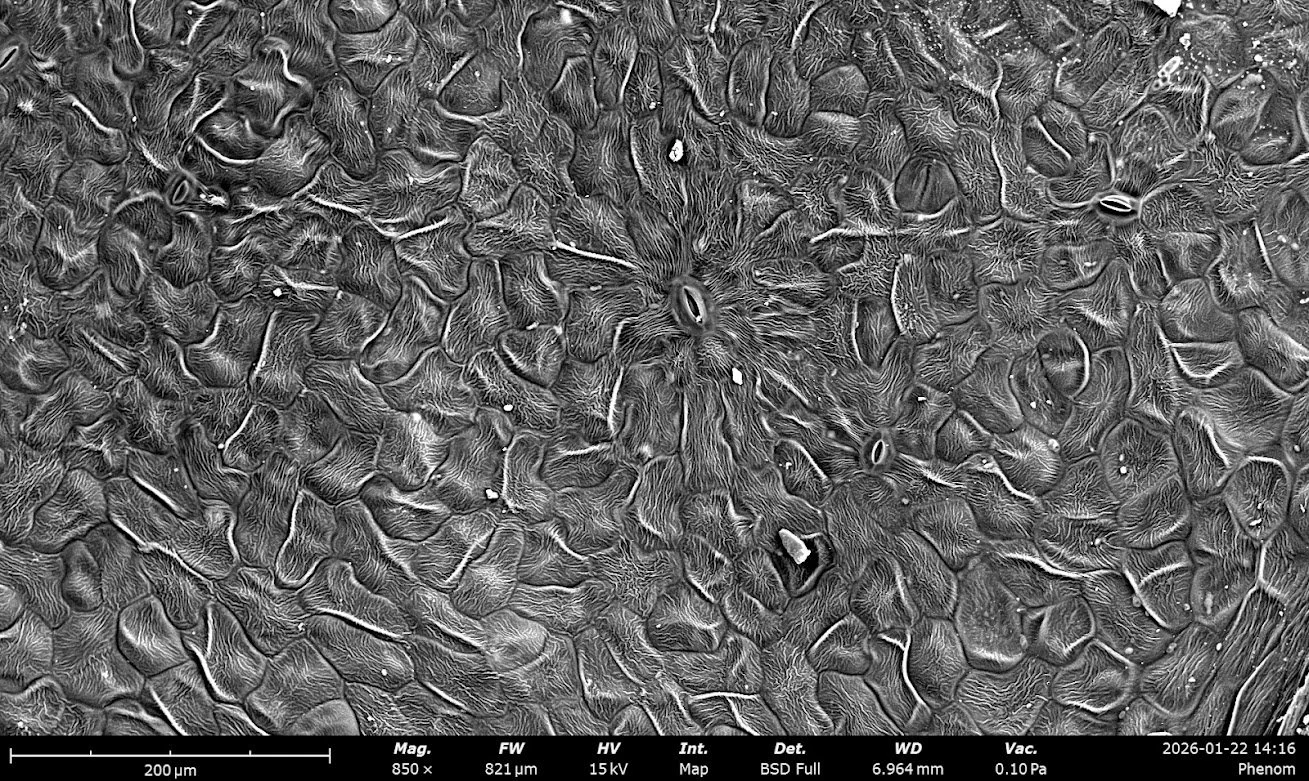
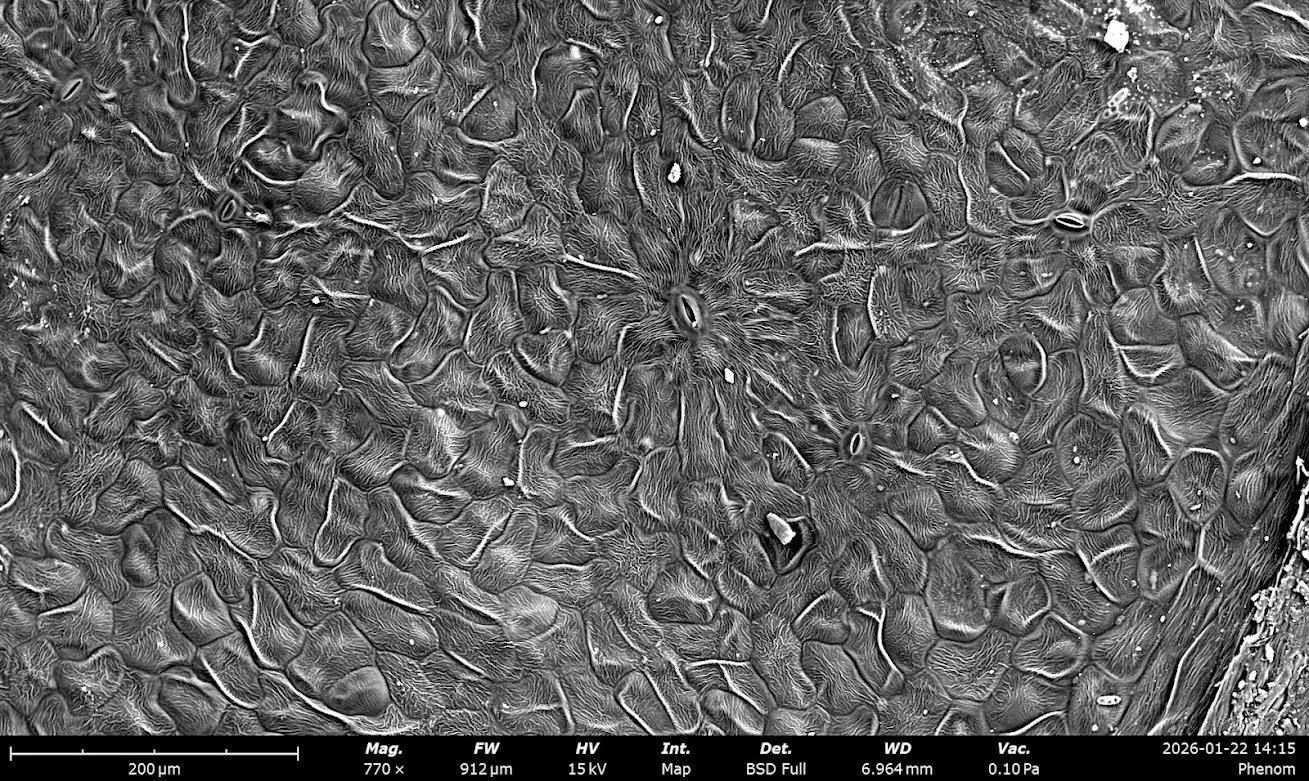
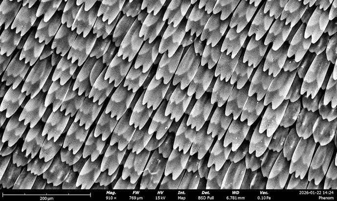
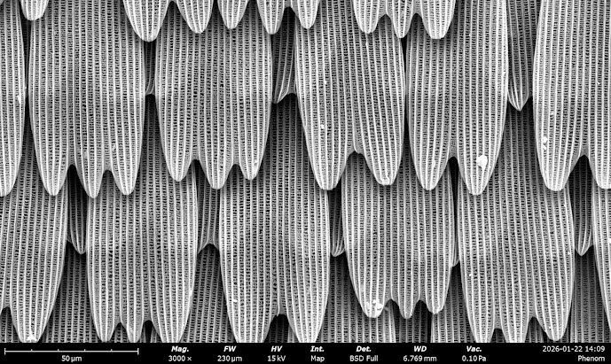
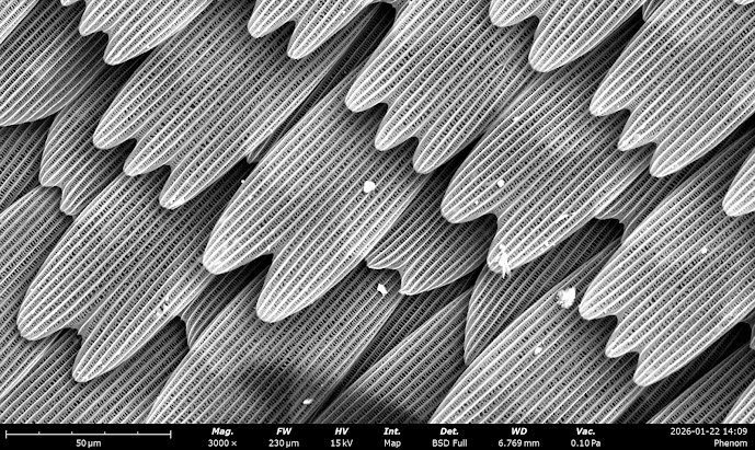
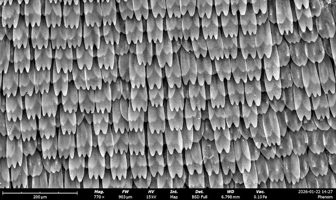
I also got trained recently on the Thermofisher Phenom XL SEM in the Breakerspace, and took photos of a butterfly wing and hydrangeas. I’m particularly interested in the forms of organic structures, which are difficult to image using SEM as it relies on samples to be dry and conductive to function. Consequently, I ensured my samples were fully desiccated beforehand, then carried out a gold sputtering process. I also had to be particularly careful when mounting my samples, as the SEM is very sensitive to particulate matter, with dust able to damage the instrument. The butterfly sample in particular was a concern as the scales can break very easily. I used a lot of compressed air to ensure the sample was clean (and definitely damaged the wing in the process), but I got great results!